Human Anatomy Rib Cage Muscles - 3D Skeletal System: Bones of the Thoracic Cage : Yet, the ribs and rib cage are also flexible enough to expand.
Human Anatomy Rib Cage Muscles - 3D Skeletal System: Bones of the Thoracic Cage : Yet, the ribs and rib cage are also flexible enough to expand.. List of skeletal muscles of the human body. The intercostal muscles extend from the. Vector art, clipart and stock vectors. This is a table of skeletal muscles of the human anatomy. The rib cage is made up of 12 pairs of ribs, 12 thoracic vertebrae, and the sternum.
Numbered ribs, sternum, cartilage parts and clavicular articulation. The pectoralis major lets you move your humerous, the minor muscle lifts your ribs, as you'll know if you are studious. Vector art, clipart and stock vectors. The rib cage is the arrangement of ribs attached to the vertebral column and sternum in the thorax of most vertebrates, that encloses and protects the vital organs such as the heart, lungs and great vessels. It, along with the skin and associated fascia and muscles, make up the thoracic wall, and provides.
Anterior and posterior view of thoracic anatomy. MVI ... from i.pinimg.com Functionally, the diaphragm separates the thoracic cavity, containing the lungs and heart and enclosed by the rib cage from the abdominal cavity, which contains the digestive. The aim of this page is to assist. This is a print of an original watercolor i made, depicting a human rib. The sternum (breast bone) is made of 3 fused bones called the manubrium, body and the xiphoid process at the very tip. Vector art, clipart and stock vectors. Human rib cage anatomy model. Welcome to the human anatomy and physiology page. Learn about human anatomy muscles with free interactive flashcards.
Human rib cage anatomy 3d model.
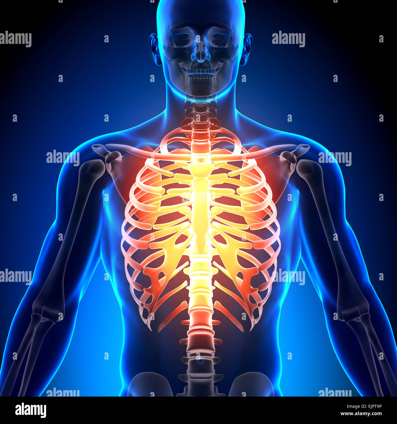
4 individual objects (spine portion, ribs, cartilages, sternum), sharing the same non overlapping uv layout map, material and uniform scale object (scale applied in blender 3d). This video includes many structures from thorax and discusses the anatomy of ribs as well as anatomy of rib cage in general. The thoracic cage, commonly called the rib cage, provides protection for the 2 lungs, heart, esophagus, diaphragm and liver. Gray's anatomy of the human body, 20th ed. If two or more fractures occur in two or more adjacent ribs, the affected area is no longer under control of the thoracic muscles. Architectural analysis of human abdominal muscles. See more ideas about anatomy, rib cage anatomy, anatomy study. • raise rib cage for inhaling & depresses rib cage for exhaling. A typical human ribcage consists of 24 ribs, the sternum, costal cartilages, and the 12 thoracic vertebrae. The rib cage, shaped in a mild cone shape and more flexible than most bone sets, is made up of varying elements such as the thoracic vertebra, 12 equally paired ribs, costal cartilage, and held together anteriorly by the sternum. Rib cage location on human body external view. Caption = the human rib cage. This is a print of an original watercolor i made, depicting a human rib.
Simplified, it is an oval that starts halfway between 1 and 2, down to mark 3; The muscle originates from the lower sternum and costal cartilage of the fifth to seventh ribs, 1 its fibers above the base of the rib cage originate from the costal margins of the ribs and terminate as (from brown shm, ward sr, cook m, et al: Vector art, clipart and stock vectors. Structure of a typical rib: They are curved and flat bones.
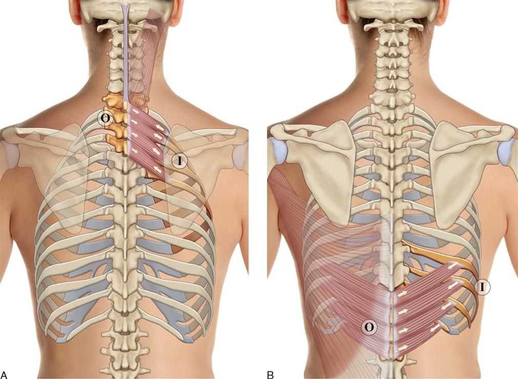
8. Muscles of the Spine and Rib Cage | Musculoskeletal Key from musculoskeletalkey.com The thoracic cage, commonly called the rib cage, provides protection for the 2 lungs, heart, esophagus, diaphragm and liver. Human rib cage anatomy model. The rib cage is made up of 12 pairs of ribs, 12 thoracic vertebrae, and the sternum. The rhomboids on your upper back let you dance your best, the minor above major. Simplified, it is an oval that starts halfway between 1 and 2, down to mark 3; The rib cage is the arrangement of ribs attached to the vertebral column and sternum in the thorax of most vertebrates, that encloses and protects the vital organs such as the heart, lungs and great vessels. Architectural analysis of human abdominal muscles. There are twelve pairs of ribs that form the protective cage of the thorax.
Human anatomy drawing drawing theory.
3d rendering medical illustration of male interior brain anatomy. Gray's anatomy of the human body, 20th ed. Almost every muscle constitutes one part of a pair of identical bilateral muscles, found on both sides. The pectoralis major lets you move your humerous, the minor muscle lifts your ribs, as you'll know if you are studious. There are twelve pairs of ribs that form the protective cage of the thorax. Caption = the human rib cage. Yet, any of these structures affected by strain, injury, or disease can cause pain. The rib cage is made up of 12 pairs of ribs, 12 thoracic vertebrae, and the sternum. The aim of this page is to assist. Learn about human anatomy muscles with free interactive flashcards. They are each attached to the ribs. Yet, the ribs and rib cage are also flexible enough to expand. The muscle originates from the lower sternum and costal cartilage of the fifth to seventh ribs, 1 its fibers above the base of the rib cage originate from the costal margins of the ribs and terminate as (from brown shm, ward sr, cook m, et al:
They are curved and flat bones. Human male anatomy, 3/4 figure muscular and skeletal systems, back and front perspective views. When you inhale, muscles between your ribs lift your ribcage helping your lungs to expand. Anteriorly, they continue as cartilage, known as costal cartilage. Human rib cage anatomy model.
Male Chest Anatomy High Resolution Stock Photography and ... from c8.alamy.com Medical human chest skeletal bone structure model. The pectoralis major lets you move your humerous, the minor muscle lifts your ribs, as you'll know if you are studious. Architectural analysis of human abdominal muscles. They are each attached to the ribs. Human rib cage anatomy model. The bones, muscle, fasciae, nerves, blood vessels and lymphatic dranaige of the thoracic wall. Human male anatomy, 3/4 figure muscular and skeletal systems, back and front perspective views. Intercostal muscles of the anterior trunk:
The muscle originates from the lower sternum and costal cartilage of the fifth to seventh ribs, 1 its fibers above the base of the rib cage originate from the costal margins of the ribs and terminate as (from brown shm, ward sr, cook m, et al:
Intercostal muscles the intercostal spaces are filled by two layers of intercostal muscles. See more ideas about anatomy, rib cage anatomy, anatomy study. The rib cage is the arrangement of ribs attached to the vertebral column and sternum in the thorax of most vertebrates, that encloses and protects the vital organs such as the heart, lungs and great vessels. Yet, the ribs and rib cage are also flexible enough to expand. This is a print of an original watercolor i made, depicting a human rib. 3d rendering medical illustration of male interior brain anatomy. It, along with the skin and associated fascia and muscles, make up the thoracic wall, and provides. Gray's anatomy of the human body, 20th ed. When you exhale, your ribcage moves down, squeezing air out of your lungs. The bones, muscle, fasciae, nerves, blood vessels and lymphatic dranaige of the thoracic wall. This is a table of skeletal muscles of the human anatomy. Learn about human anatomy muscles with free interactive flashcards. Almost every muscle constitutes one part of a pair of identical bilateral muscles, found on both sides.
The pectoralis major lets you move your humerous, the minor muscle lifts your ribs, as you'll know if you are studious rib cage muscles. Gray's anatomy of the human body, 20th ed.